Opportunities of Connectomic Neuromodulation

Clinician Scientist, Harvard Medical School
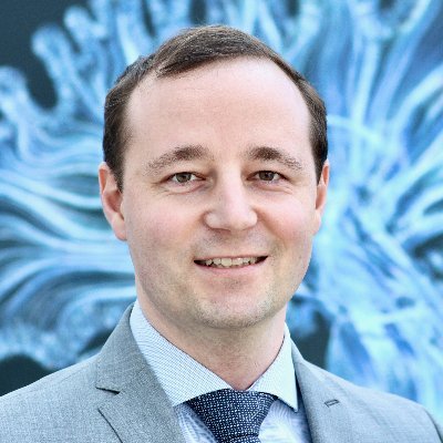
Professor Andreas Horn first studied medicine in Freiburg and during his MD thesis became interested in neuroimaging. He later went to Berlin to pursue his career and focused on connectomics. He was using non-invasive methods like functional MRI and imaging-based tractography to study non-invasively the broader connections in the brain of living humans. He was fortunate enough to be able to join Andrea Kühn’s group at the Charité Berlin where he was also involved in Deep Brain Stimulation localization. He thought it would be great to combine the two fields and focused some of his work on making precise reconstructions of where exactly the electrodes would line up in the brain. That coincided with him going to Harvard for a postdoc with Prof. Michael D. Fox. They were researching what differentiates the patients that would have great motor improvement after surgery in Parkinson’s Disease. He later went back to Berlin and founded his lab in 2018 and now he has been recruited back in Boston as a faculty member at the Brigham and Women’s Hospital with Prof. Michael D. Fox.
What is the brain's "connectome"?
The connectome term is a great term –or was a great term from Olaf Sporns in 2005- and now I think is used a bit as a hype thing. We have to differentiate brain connectivity, which goes back to the 1920s when anatomists looked at networks and treated Parkinson’s Disease as a network disorder since that time or probably earlier. So not at all new, brain connectivity means two things are connected in the brain and they form a network and a circuit. Even back then people thought about modulating networks.
The connectome term is new and was originated in the imaging world and essentially means that we break up the whole brain into parcels and then look at their interconnections. Often that’s visualized by a mathematical structure. That has powerful consequences, you could look at specific properties of that graph, for example how modular or central each edge or node of it is to the network. That’s really the connectome versus connectivity being a much broader term that just looks at brain connectivity. I’d say that most of what we and other people do in the DBS field currently, is connectivity DBS, it’s not connectome DBS. However, people have looked at it in that connectome fashion, for example, how the centrality of a node declines with DBS. That would be true connectomic DBS. My lab is looking a lot in connectivity. There are some things that widely overlap with the connectome fields in the imaging domain. For example, we sometimes look at similarities of whole-brain connection profiles, which could deserve this connectome term. However, I think that the more important thing is which connections are crucial for clinical outcomes.
Why do we need connectomics for Deep Brain Stimulation?
There are two levels we could look at it. The local level, meaning where to stimulate (the sweet spot), what coordinates to use and which network to target. We can explain many variants based on the local level and say “Stimulating this spot is great! Why do we need connectomics?”. There are a few reasons.
One would be more from a basic science perspective, it’s really helpful to know which networks are associated with that sweet spot. It’s not really that we need to modulate the STN but we also need to modulate its connection with the pre-motor cortex. So that will help us better understand Parkinson’s Disease or Obsessive-Compulsive Disorder as diseases for example. We can use DBS as a tool to investigate the brain’s functional connectome. Because if you have such a graph and you can modulate one node of it, you can see what happens in the network. That’s a really powerful tool for basic science reasons.
Now for clinical reasons, there are also great examples where the connectomic part really shined. For example, we could show in Obsessive-Compulsive Disorder that it’s a disease from the neuropsychiatric domain. There are different target sites that have been discussed that would need to be stimulated. One is the subthalamic nucleus and another is the anterior limb of the internal capsule. We could show that the same tract that connected the two was important to modulate both of these targets. We could even use connectomics to cross-predict across the two targets, then built a model of optimal connections just based on one target. One cohort operated with that one target and then predicted the ranks of the other arm of the other target. That was maybe the best demonstration so far for why connectivity could matter because we could not only show that this is the spot for one target, but we could show that this would be the network and maybe you can stimulate it at different sites.
What impact can connectomics and adaptive Deep Brain Stimulation (aDBS) have in patients undergoing neuromodulation?
Jonathan: It really is true that we very frequently still see patients that have a poor outcome from DBS in OCD but in movement disorders as well. Usually, in the videos, they never show the patient that had a bad response, they always show the one who had a miraculous recovery of function.
Andreas: You’re totally right. Some people already do great why do we even need to investigate it more? However, I think right now probably in Parkinson’s DBS -just gut feeling- 90% will respond, but only let’s say 30% will be these excellent responders and my aim is to make that 30%, 90% or at least try. That could be done with connectomics but also just with good imaging. So it’s both levels that are important, the local one but also the network level and trying to get more deliberate. We probably won’t be able to improve the 30% even further with what I’m doing, but we could make more people top responders.
Jonathan: Do you think, clinically speaking, those 30% are getting as good a response as it’s possible to get?
Andreas: I would think as good as possible with what we’re currently doing, with this classical 130 hertz always on DBS. To improve those further, we need something new, disruptive, which could be adaptive DBS. So far it doesn’t look like patients would really get better than with continuous. Adaptive DBS is when the system listens to the brain and then modulates based on specific rules.
Jonathan: We’ve seen that with the release of the first commercial device, the Medtronic percept system. So that’s a reality now, but you’re saying that we’ve yet to see real evidence that that’s the game-changer or that it’s going to be the game-changer.
Andreas: I would hypothesize that it won’t change these top responders even further in terms of their motor outcome, but it could maybe help reduce side effects. The fact that the system isn’t always on could mean that it’s only on when really that effect is needed, but then it’s often maybe not detrimental to some other function like speech if it’s adaptively off when not needed. I think with the current technology, it will be hard to improve these ultimate top responders because they often already go down to nearly no symptoms anymore for a while and then Parkinson’s would progress further, unfortunately. That is probably the nature of the disease, it’s neurodegenerative so we can’t restore that with DBS at the moment.
What are the two main modalities of connectomics in neuromodulation?
Andreas: One is functional MRI, an indirect method of brain activity that uses what’s called the bold contrast, the blood oxygenated level-dependent contrast. It essentially uses the fact that haemoglobin is slightly different in terms of its magnetic properties whether it’s oxygenated or not. It uses brain oxygen and blood flow ratios to derive which area is active in the brain. So if you have a human seeing a flicker board of light then the v1 reach in the primary visual cortex will light up and we will have more of that bold signal there. People have come up with good risk resources over the years.
Now resting-state function MRI would be to look if the two parts of motor court disease would co-fluctuate and if their signal would go up and down in the same way they would be just correlated in time. That is what we call functionally connected. It’s very far from a real axonal type of connection, it is more a statistical rough guess that these regions do something together, but what they do together we don’t know. So the temporal resolution is also really slow in fMRI. It all boils down to that it’s a poor man’s version of connectivity but it works in living humans and we can use that to estimate things, which is amazing. It’s a very derived method.
My lab, rather than using patient-specific data, uses big cohorts of scanned brains, plus histology data or more textbook connectivity data to infer with the patients. We know where the electrode is, we know where the structures are and in that way, we know what they are usually connected to when we exploit it. We’ve done both, we’ve looked at patient-specific data but also more high-resolution atlas data.
Jonathan: Is it possible to combine the two forms of data in the same patient?
Andreas: I would love to do that! That’s one of my focuses for the next year, to find good ways of emerging information from a high resolution -even postmortem- connectome that was scanned in the best centre in the world, probably here in Boston Martino Center, also with the patient-specific scans and then individualize the high-resolution data. So I think that’s definitely a fruitful future topic, so then you have the best of both worlds and a patient-specific high-resolution connectome.
What would clinical implementation for connectomic neuromodulation look like?
Lots of great ideas! Mike Fox has just published one paper in brain looking at networks that would impact cognitive decline based on Deep Brain Stimulation. A few years ago, we also had a paper and annals that looked at which connections lead to depression in Parkinson’s Disease.
One avenue that we’re trying to investigate is looking at our database locally and the patients who might have that symptom and reinviting them to the centre and trying to reprogram them. I think in the long-term my vision would be to have symptom-specific network profiles, let’s say one for tremor, one for bradykinesia, one for rigidity and then also for depression and cognitive decline and all that mapped out in a normal brain as a library. They are not patient-specific, but they are maps that we can use and if a new patient comes in we could check their symptoms scores. Let’s say they have a lot of tremor, we would then weigh the tremor network more strongly to plan their surgery and then also to program them. So for each patient we could weigh the profiles we have and find their optimal mix or blend of networks, call that network blending, and then based on that do surgery but also DBS programming. I think that will be the future. There will be many other things in the future as well, like adaptive DBS and so on, but I think this will certainly evolve to be ripe for clinical practice.
An intermediate step that we haven’t talked about is that if we have these libraries and patients’ symptoms, we would also want to scan their brains and match them to the library networks. In that way, we would see it in the individual patient. So we would scan them before surgery, they would get a resting-state fMRI and a GPI scan and then we would segregate their brain into the networks which we know would respond to tremor or bradykinesia for example and then do the network blending in their own brain and of course have it all individualized.
This is the highlight of the interview. If you like to explore more, please visit our YouTube Channel.


